.webp)
Impacted wisdom teeth with multiple risk factors
Many wisdom tooth complications centre on nerve proximity. That is a real and serious risk, but it is not the only factor that makes a case complex. Root morphology, bone density, and anatomical variants each carry their own surgical implications. In older patients, these factors tend to arrive together.
A 41-year-old male presented with long-standing pain associated with all three wisdom teeth. The lower teeth were causing the most significant discomfort and had produced recurring symptoms over time.
The lower wisdom teeth were vertically impacted, positioned below the gumline with the crown facing upward, but vertical impaction does not necessarily mean straightforward removal. Initial examination indicated several features that warranted a closer look before surgical planning could begin.
Pre-operative imaging
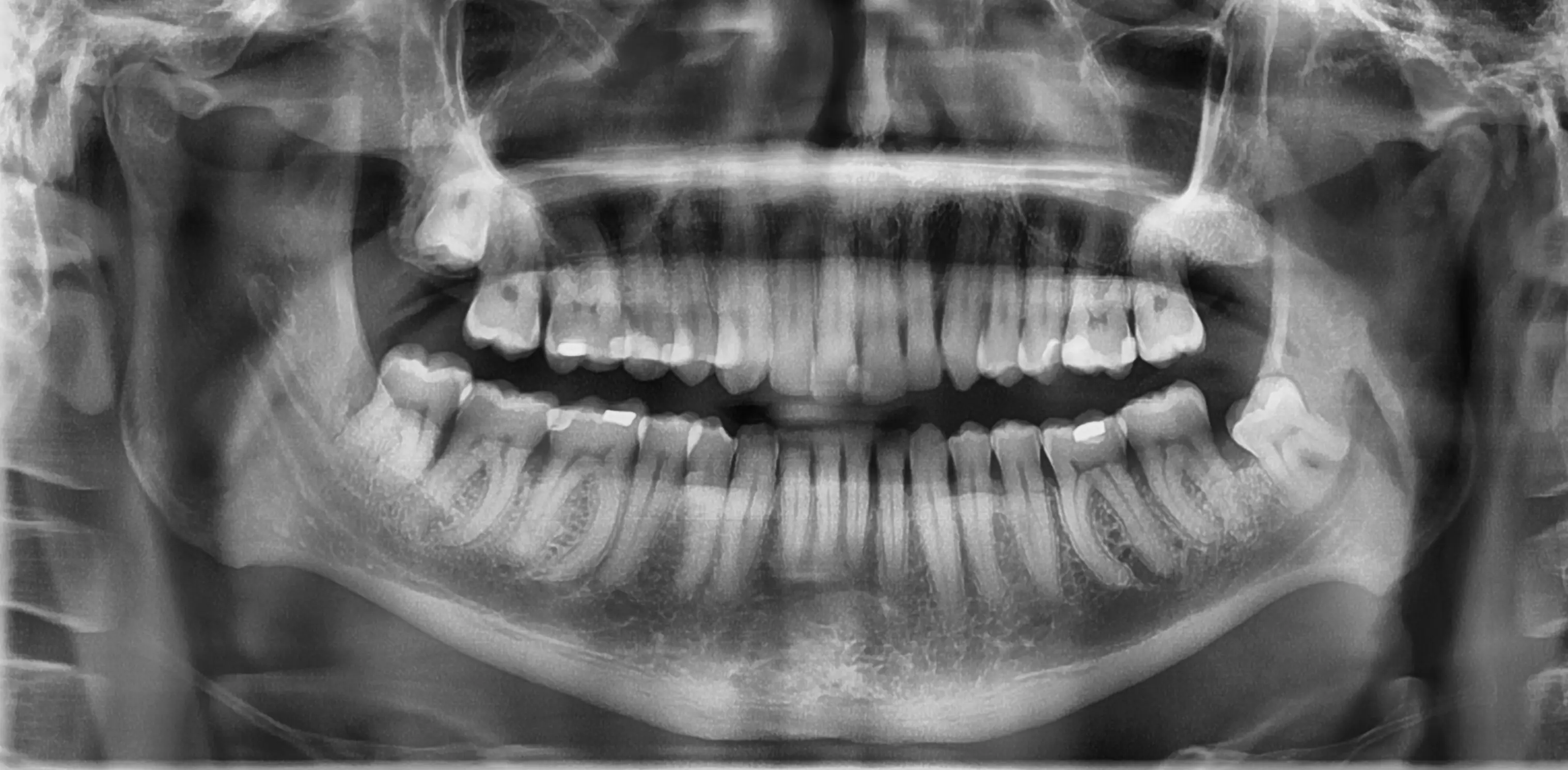
An initial Orthopantomogram (OPG) was taken as the first step in the surgical planning process. The OPG provided a useful overview of the position and general morphology of all wisdom teeth, but it is a two-dimensional image of a three-dimensional structure, so fine detail about root shape or the precise relationship between roots and nerves is often obscured. A Cone Beam CT scan (CBCT) was ordered for a more detailed assessment.
Surgical approach and outcome
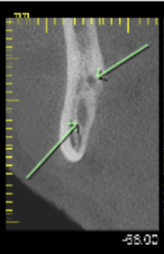
Given the range and severity of the complicating factors, removal under general anaesthetic in a hospital setting was the appropriate choice. Several factors contributed to this, including the age of the patient, unusually bulbous roots, areas of hypercementosis, and the very close proximity of the lower wisdom teeth to the inferior alveolar nerves (IAN). The CBCT scan also showed a second branch of the right IAN, adding a further layer of complexity, along with the upper right wisdom tooth positioned close to the maxillary sinus.
Age is a relevant factor in wisdom tooth surgery. As we age, the jawbone naturally becomes harder and denser, and wisdom teeth often seem to "fuse" to the surrounding bone. Although the lower wisdom teeth were vertically impacted in this case, their removal was not straightforward and required careful sectioning of the teeth to minimise trauma and protect the surrounding structures.
Both lower teeth were removed with particular attention to the nerve anatomy identified on the CBCT, including the right-sided duplicated IAN. The upper right tooth was removed with care given its proximity to the maxillary sinus. The patient recovered well without complications.
This case highlights the importance of thorough planning when managing complex wisdom teeth. When wisdom tooth roots are positioned close to important structures such as the IAN, advanced imaging with a CBCT scan provides invaluable information that cannot be seen on a standard dental X-ray alone. This allows treatment to be planned more precisely, helping to minimise surgical risks and optimise patient outcomes.
When to refer wisdom tooth cases
Certain presentations of wisdom teeth issues fall outside the scope of what can be safely managed in a routine dental setting. Referral to an Oral and Maxillofacial Surgeon is appropriate when imaging shows increased risk factors. Complex wisdom tooth extractions, particularly those involving close nerve relationships, are often best managed by an experienced Oral & Maxillofacial Surgeon. In many cases, treatment under general anaesthetic in a hospital setting offers the safest and most comfortable experience for the patient. Anatomical variants such as a duplicated IAN, as seen in this case, are not predictable without advanced imaging and require a surgical approach that accounts for the variant.
Dr Franc Henze at Western OMS accepts referrals for complex and high-risk wisdom tooth cases across Perth and is always happy to assist with assessment, advanced imaging, and surgical management.
.webp)